![]()
Colonies of cells enclosed in a mucilaginous sheath
page in preparation
| spherical/elliptical mucilage sheaths | ||||
| (a) with flagella or pseudocilia | ||||
| (i)
pseudocilia (non-motile hair-like appendages) |
||||
|
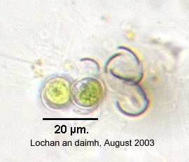
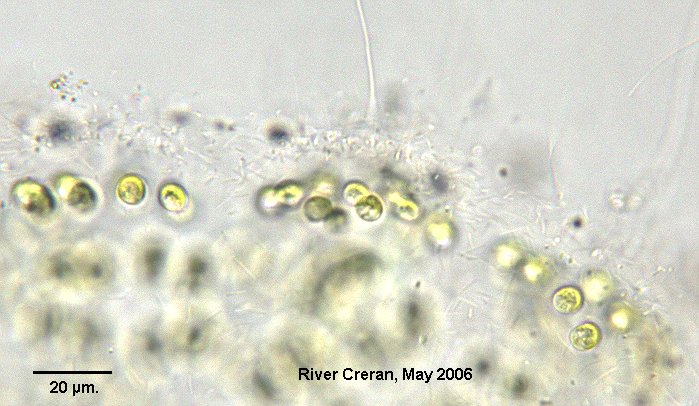
Apiocystis brauniana |
Cells around the periphery of a sac-like mucilaginous envelope, attached at one end. Pseudocilia project through the mucilage | 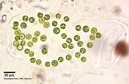 |
||
|
Tetraspora
|
 |
|||
| (ii) flagella: motile appendages which make the colony motile | ||||
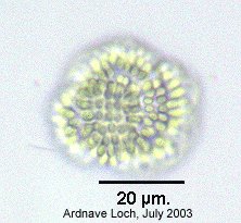
|
Eudorina
FAFBI p. 317 plate 81 |
each cell has a pair of flagella; cells spaced out from each other |
 |
 |
 |
|
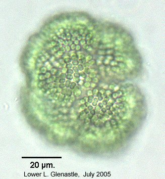
Pandorina
FAFBI p. 320 plate 81 |
each cell has a pair of flagella; cells tightly packed together; angular from mutual compression | 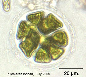 (Lugol's) |
 |
|
| (b) without flagella or pseudocilia | ||||
| (i) Cyanobacteria: cells with chlorophyll not enclosed in a chloroplast (not always easy to see) | ||||
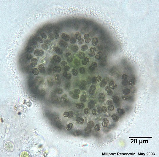
|
Coelosphaerium
FAFBI p.42 plate 5 |
Colony spherical or subspherical, the cells grouped around the outside forming a hollow ball | |||
|
Woronichinia
FAFBI p.58 plate 5 |
Similar to Coelosphaerium but cells are somewhat elongate, at the end of radiating stalks
|
 |
 |
|
|
Snowella
FAFBI p.56 plate 4 |
|
 |
||
|
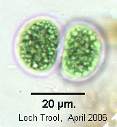
Chroococcus
FAFBI p.40 plate 3 |
Cells in pairs or multiples of 2, somewhat flattened where the cells are in close contact (sometimes with concentrically striated mucilage). |  |
 |
|
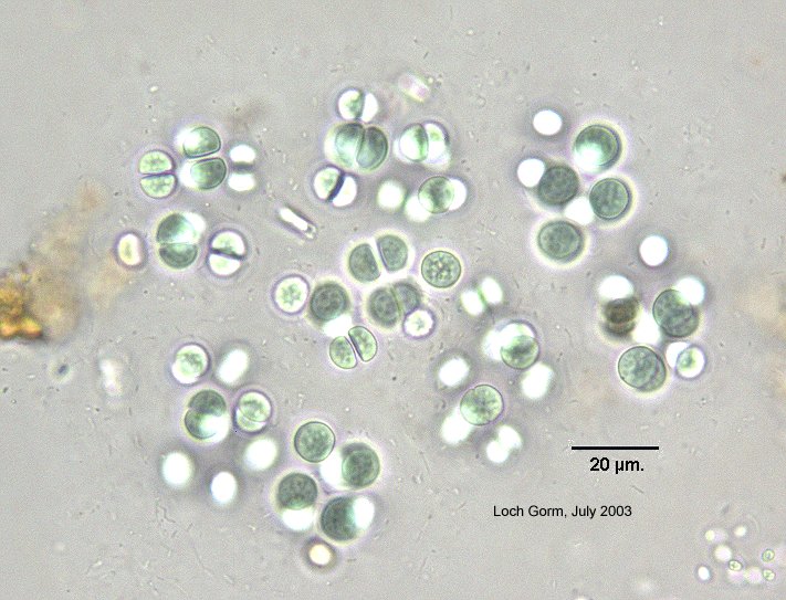 C. limneticus |
 |
|||
|
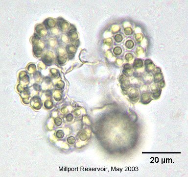
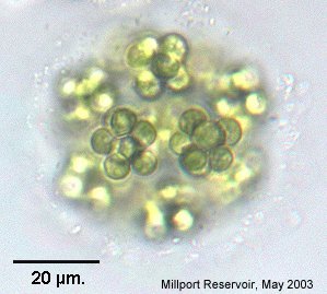
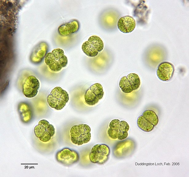
Eucapsis
FAFBI p.45 plate 5 |
Cells in groups of multiples of 4 in more or less cubical arrangement | 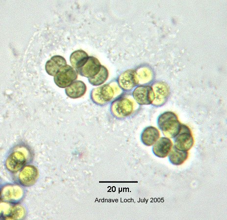
 |
 |
|
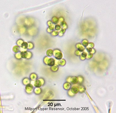
|
Merismopedia
FAFBI p.51 plate 3 |
Cells in groups of 4 or multiples of 4, forming a flat (or sometimes rolled) plate. | 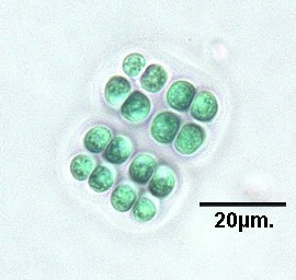 |
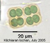 |
|
|
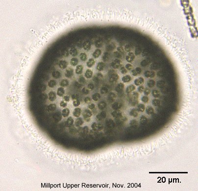
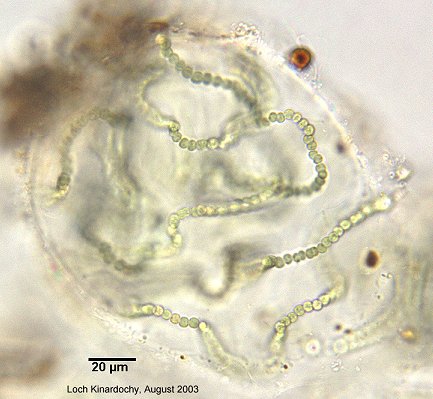
Nostoc
FAFBI p.105 plate 18 |
Clusters of filaments in a firm mucilaginous envelope. The cells are spherical with occasional heterocysts (rather similar to Anabaena) | 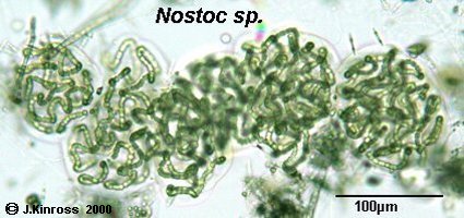 |
 |
|
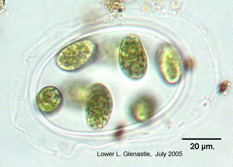
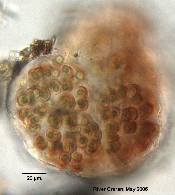
| (ii) cells with chloroplasts, in groups within a concentrically layered mucilage | ||||
|
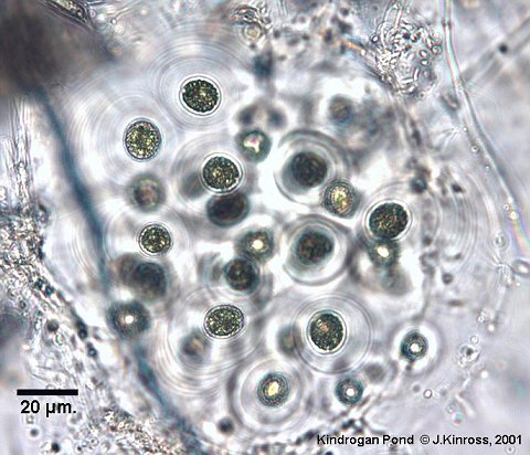
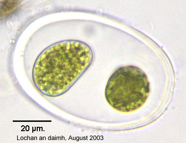
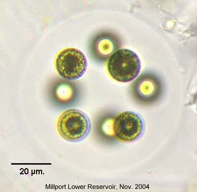
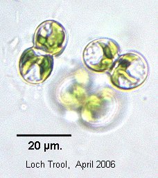
?Gloeocystis
FAFBI p. 356 plate 86 |
 |
 |
 |
|
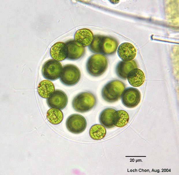
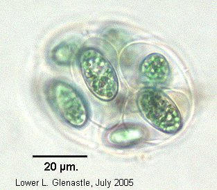
|
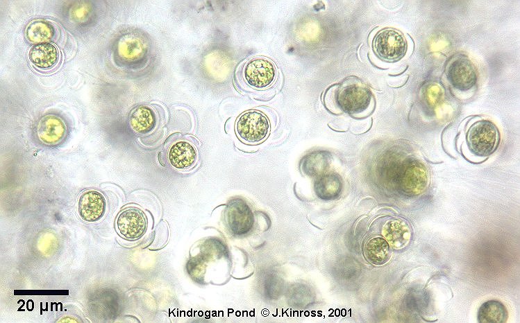
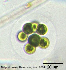
Asterococcus
FAFBI p.299 plate 76 |
Cells
spherical or subspherical, with stellate chloroplast. Mucilage envelope
wider than cell. |
 |
 |
|
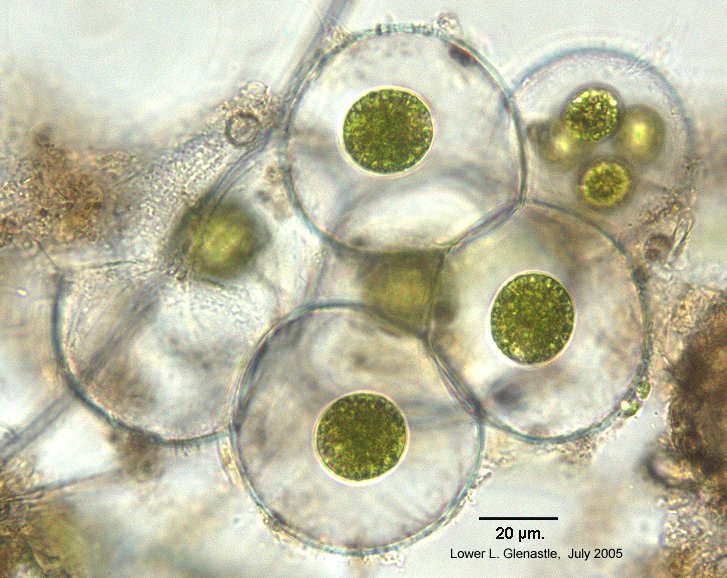 |
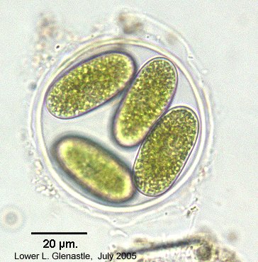
||||
| Non-lamellate mucilage, cells 10-25µm = A. limneticus |
 |
 |
||
|
Lamellate mucilage, cells 30-43µm = A. superbus |

|
 |
||
|
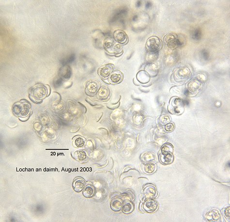
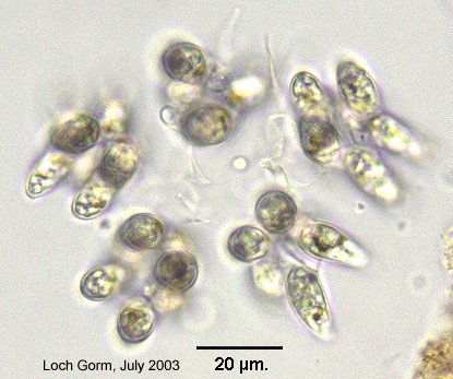
?Oonephris
|
 |
|||
|
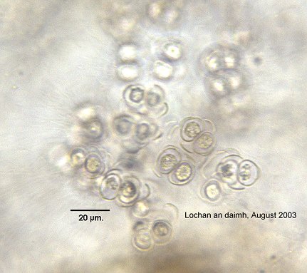
Schizochlamydella
FAFBI p.398 plate 86 |
 |
|||
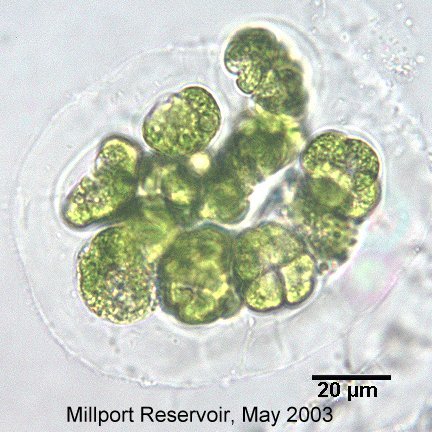
|
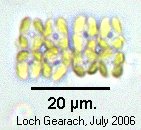
Westella
FAFBI p.408 plate 84 |
 |
 |
 |
|
|
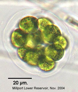
?Oocystis
FAFBI p.372 plate 92 |
 |
 |
 |
|
|
?Oocystis borgei |
 |
 |
||
|
?Coenococcus
polycoccus
FAFBI p. 343 plate 86 |
|
 |
||
 |
 |
|||
|
?Nephrocytium
limneticum
FAFBI p.371 plate 83 |
|
 |
||
|
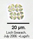
?Coelastrum
sphaericum
FAFBI p.342 plate 83 |
 |
 |
||
| Unknown species to match | 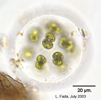 |
 |
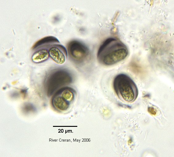 |
 |
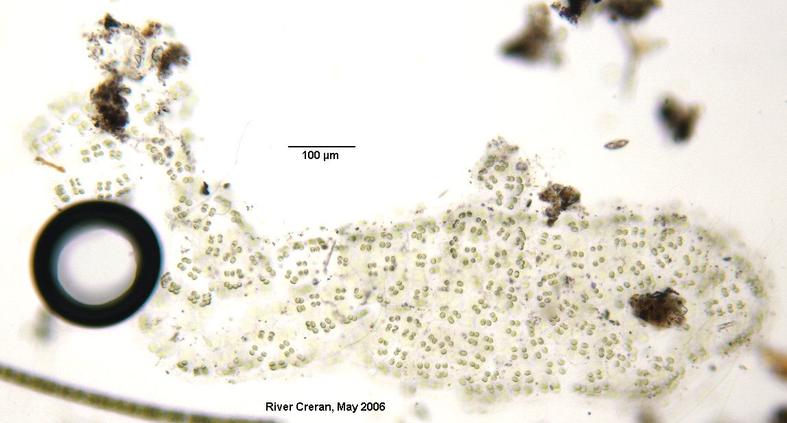 |
 |
 |
||
|
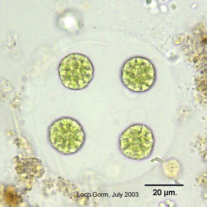
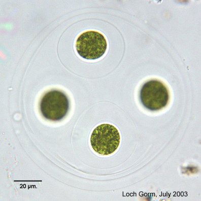
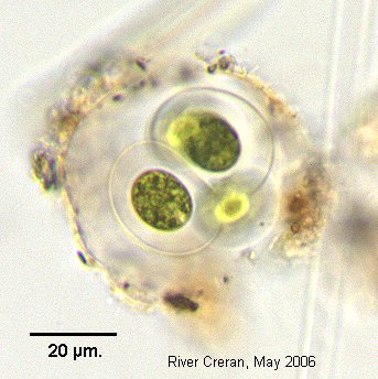
?Chlamydocapsa
FAFBI p.300: dubious genus, not illustrated (may be identical to Chlamydomonas palmelloid stage) |
 |
|||
|
Chlamydomonas
palmelloid stage
FAFBI p.308 plate 77 |
||||
|
?Coenocystis
obtusa
FAFBI p.343 plate 86 |
 |
|||
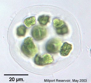
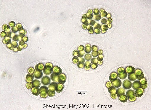
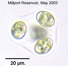
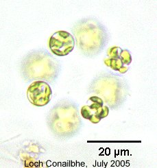
|
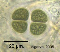
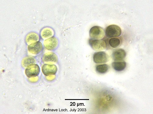
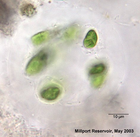
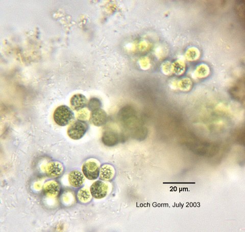
Sphaerocystis
FAFBI p.402 plate 86 |
 |
 |
||
|
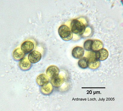
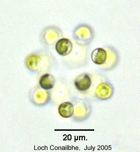
?Sphaerocystis
FAFBI p.402 plate 99 |
 |
 |
||
|
Cosmocladium
saxonicum
FAFBI p.550 plate 143 |
 |
 |
 |
 |
|
Glaucocystis
FAFBI p.613 plate 1 |
 |
|||
|
?Dichotomococcus
FAFBI p.347 plate 84 |
 |
|||
|
?Tetrastrum
FAFBI p.406 plate 94 |
 |
|||
|
Crucigenia
FAFBI p.344 plate 84 |
 |
|||
|
Crucigeniella
FAFBI p.406 plate 84 |
 |
|||
|
Quadrigula
closterioides
FAFBI p.382 plate 98 |
 |
 |
||
|
?Rayssiella
Not present in FAFBI; ID from Prescott |
||||
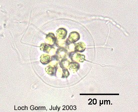
|
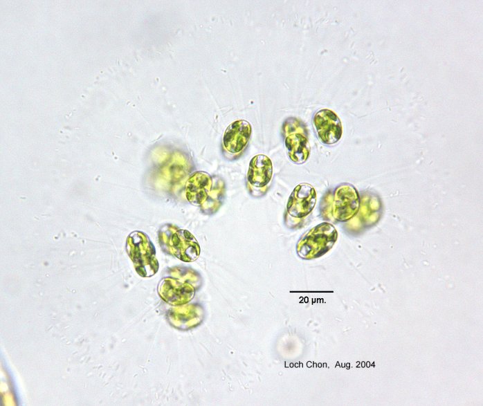
?Palmodictyon
FAFBI p.375 plate 99 |
colonial Chlorophyte: spherical cells in branching tubes of mucilage, with cup-shaped chloroplast | 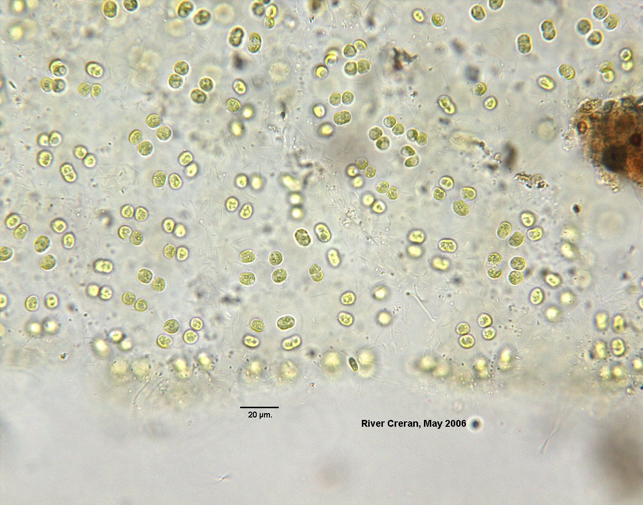 |
 |
|
|
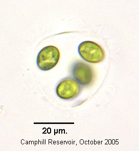
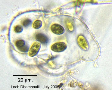
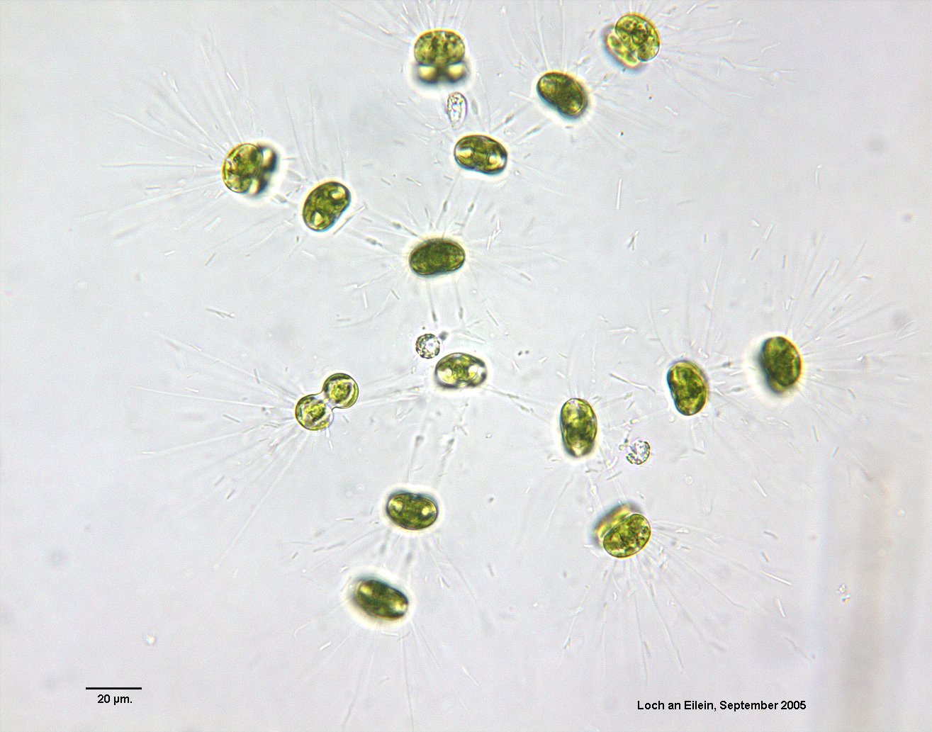
?Paulschulzia
FAFBI p.300 plate 76 |
colonial Chlorophyte:cells/cell groups in clearly defined spherical mucilage envelope, with individual sheaths and flagellum extending beyond outer envelope | |
 |
|
|
?Phaeosphaeria
FAFBI p.234 plate 62 |
 |
|||
|
?Radiococcus
FAFBI p.383 plate 86 |
 |
|||
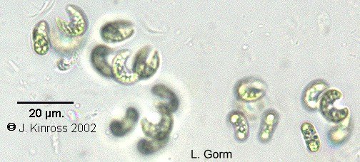
| Sheath not spherical | ||||
|
Raphidocelis
(Kirchneriella) FAFBI p.383 plates 83, 98 |
 |
|||
|
Schizochlamydella
FAFBI p.398 plate 86 |
|
|||
|
Westella
FAFBI p.408 plate 84 |
|
|
|
|
|
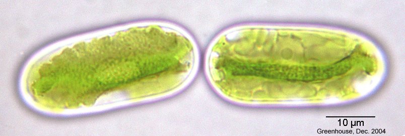
Mesotaenium
FAFBI p.511 plate 128 |
 |
|||
|
|
||||
![]()
![]()
John Kinross